
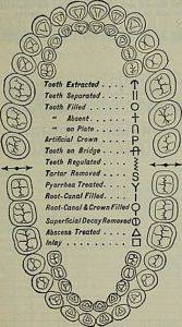
A odontogram, Also called dentogram, periodontogram, dental chart or dental diagram, it is a diagram of the dental arches. It shows graphically and anatomically all the teeth. In this scheme, the dentist indicates the various pathologies and treatments with a color code.
Numbers are generally used to identify each tooth, however teeth can sometimes be numbered with capital letters or number pairs. In the diagram, both the primary (children) and the definitive dentition are marked.
The odontogram is part of the dental clinical history of a patient, since it gathers all the information related to the mouth, the anatomical characteristics of the teeth and other peculiarities, indicating the treatments required, the follow-up, etc..
It is a fundamental tool for the diagnosis, treatment and monitoring of a patient's dental pathologies. The odontogram is also used by forensic dentistry for the identification of cadavers, since the teeth are very resistant and do not decompose like other organic tissues..
The odontograms are filled out physically, that is, on a printed sheet; however, there are currently many computerized programs that allow the digital record to be maintained. This facilitates the recording of information and the storage of medical records..
The digital registry allows information to be shared quickly and easily and to make inter-consultations between specialists in the area without initially mobilizing the patient.
Article index
It is an important part of a patient's medical history and, in its content, the dentist captures information related to:
-Patient identification
-The general condition of the oral mucosa
-What teeth to treat
-The treatments the patient has had previously
-Patient follow-up
-Temporary and permanent dentition in children
The odontogram facilitates the exchange of information between dentists for the different consultations. Due to its importance it is necessary to update it frequently, preferably with each patient visit.
The odontogram is the equivalent of a patient's medical history, it preserves information regarding all dental pathologies, their treatments and evolution. Currently that information is stored digitally.
There are two types of odontograms: one anatomical and the other geometric. Although any odontogram can be customized, each type follows a particular methodology.
It uses a representation of the exact anatomical shape of the different teeth. There are four types of anatomical odontograms which are:
-Walter Drum diagram: also called the FDI system, it uses two digits, one to define the quadrant or position and the other to identify the tooth.
-Sign diagram: uses a negative sign for the lower arch teeth and a positive sign for the upper arch.
-Numerical diagram: also called a universal diagram because it is one of the most used. Each tooth has a number, the first upper right molar has the number 1, the last one is the third lower right molar, with the number 32.
-Zsigmondy diagram: divide the oral cavity into quadrants and identify each part with a number.
Makes a representation using geometric shapes, such as circles and squares, to represent each of the faces of each tooth
The International Dental Federation, with the approval of the World Health Organization, proposes a nomenclature that includes two numbers for each tooth.
The first number refers to the quadrant where the tooth is located. The dental arches are divided into four quadrants that are numbered from 1 to 4.
The second number indicates the position of the tooth in the respective quadrant. There are eight permanent teeth or five temporary teeth (children up to six years of age) that make up each quadrant.

Following the interincisal midline, the dental arches are divided into four quadrants: two upper (one right and one left), and two lower (one right and one left). The numbering of the quadrants is as follows:
The second digit places the tooth within the quadrant that is called hemi-arch or semi-arch. Teeth are numbered from the midline backward from 1 to 8.
For primary or primary teeth, the first number refers to the quadrant with the same division described above, but with the numbers from 5 to 8 as follows:
The teeth in each quadrant are identified from 1 to 5 as follows:
There are some letters that are used to identify the face of the tooth as follows:
V = vestibular
M = mesial
D = distal
P = palatal
L = lingual
O = occlusal
Linear symbols and colors are used to identify dental lesions or pathologies. The colors used are red, blue, orange and green. Lines are horizontal, zigzag, oblique, or circular.
The color red is used to locate dental cavities on the tooth and on the corresponding face. Blue is used when the treatment is in good condition. Green for temporary treatments. Orange color is used for resins.
Each tooth has a geometric representation of its faces. Upwards is the buccal face, downwards the palatal or lingual, if it is of the upper or lower arch respectively, the central square of each tooth represents the occlusal face and the right and left square represents the distal and mesial face..
Due to the length (more than 20 symbols and specific acronyms), only a few symbols will be named as representative examples..
- The cavities are painted red, located in the entire extension of the compromised dental faces, trying to imitate the shape and extension.
- An oblique blue line, passing through the drawing of a tooth, indicates a missing tooth.
- Two horizontal red lines on all representations of the faces of a tooth indicate root remnant.
- Some areas of different dental faces painted green indicate temporary fillings.
- Some areas of different tooth surfaces painted blue indicate fillings with amalgam. The abbreviation ME is placed in the upper box if it is in poor condition.
- Some areas of different dental faces painted orange indicate fillings with resin. The abbreviation ME is placed in the upper box if it is in poor condition.


Yet No Comments