
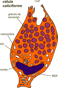
The goblet cells they are secretory cells or single-celled glands that make and expel mucus or mucus. They receive that name because they are shaped like a chalice or cup. The upper part of these cells is wider - cup-shaped, where secretion vesicles are stored - and the lower part is a narrow base, like a stem, where the nucleus is located..
These cells are widely distributed in the epithelium or tissue that covers many organs. They are found mainly in the respiratory system, in the trachea, bronchi and bronchioles, in the conjunctiva membrane of the eyes and in the intestines, being there where they are most abundant.
When the goblet cells release the produced mucus, they decrease in size and begin to store it again. This is how they make secretion cycles, in which they fill and empty every 1 or 2 hours.
Goblet cells and the mucus they produce have been little appreciated and researched. More detailed studies are required to better understand the work of this cell, its contribution in immunology and in the balance in the functions of the organs.
This study can also be valuable in the design of new treatments for many diseases associated with these cells..
Article index
Goblet cells, also known as goblet cells by their English name, are goblet-shaped cells that have the function of secreting mucin.
Mucin is a mucopolysaccharide, a normally translucent and viscous material that dissolves in water to form mucus..
This mucus is mainly a lubricant: it prevents dehydration of the mucosa, protects against infections and diseases, and is a stabilizer of the flora in certain organs.
Goblet cells were first observed and named by German scientists. The first to notice them was the doctor Friedrich Gustav Jakob Henle in 1837, who identified them in the mucous membrane of the small intestine..
It was not until 1857 that the zoologist Franz Leydig called them mucous cells, after examining the epidermis of fish.
In 1867 Franz Eilhard Schulze (also a German anatomist) gave them the name goblet based on their shape, as he was not sure that these cells secrete mucus.
These cells synthesize mucinogen (name of the substance inside the cell) or mucin (name outside the cell). Mucin release occurs by merocrine secretion; that is, during the secretion process there is no presence of any type of lesion in the secretory cell.
Mucus secretion is preceded by a stimulus. Together with the secretory granules, they secrete mucus through exocytosis (process in which the contents of the vacuole are released).
Goblet cells have a very outstanding morphology: mitochondria, the nucleus, the Golgi body and the endoplasmic reticulum stand out in the basal portion of the cell (an extracellular section composed of proteins). The rest of the cell fills with mucus into secretory granules.
Regardless of whether they accumulate mucus or not, the shape of goblet cells always changes. This is how young cells are rounded, and they flatten and increase in size with the passage of time..
Disseminations are found between the epithelial cells that line the small and large intestines; in the respiratory system, trachea, bronchioles and bronchi; and in certain lubricated epithelia.
These cells associate to form groups called intraepithelial glands, which can be found in the nasal cavities, in the Eustachian tube, in the urethra and in the conjunctiva of the eye, where they provide mucin secretion together with the Manz glands, forming a mucous layer or tear film.
In addition to forming the epithelial lining of various organs, goblet cells produce carbohydrates and glycoproteins, but their most significant function is the secretion of mucus..
Mucus is a viscous substance that is composed mainly of mucins, carbohydrates, and lycoproteins..
Its function in the small intestine is to neutralize the acids produced by the stomach and lubricate the epithelium, to facilitate the passage of food.
In the large intestine, the mucus layer formed prevents inflammation, since it prevents the passage of bacteria derived from food that pass through it.
In the respiratory tract, they trap and carry inhaled foreign bodies; this is where they produce more mucus than in any other part of the body.
They also perform functions in the conjunctiva of the eyes. The conjunctiva is the thin membrane that covers the exposed areas of the eyeballs and the inner area of the eyelids..
These organs, which are in contact with the outside environment, are lined with goblet cells that, together with the secretion of tears, function for lubrication and against foreign agents..
Just as goblet cells can perform a beneficial work for the body, an excessive proliferation of them (or hyperplasia) can be harmful.
It is also detrimental when these cells undergo metaplasia; that is, when they change, becoming another type of cells.
Efficient mucus flushing helps keep lungs healthy. If there is an excessive increase in the production of mucus, it cannot be eliminated and obstructs the airway, causing difficulty in air flow and favoring the colonization of bacteria..
The mucociliary defense mechanism is essential to maintain sterility in the airways. Changes in the mucociliary sweep contribute to the generation of infections and the development of respiratory diseases, such as COPD and asthma.
To treat these diseases there are various mucoactive compounds, such as expectorants, mucoregulators, mucokinetics and mucolytics..
An example of alterations in the case of the digestive system would be the so-called Barrett's esophagus. The lining of the esophagus has squamous cells. Goblet cells are normal in the intestine, but not in the esophagus.
It is said that there is intestinal metaplasia when goblet cells grow in a place where it is not normal for them to do so; in this case, the esophagus.
Barrett's esophagus occurs when the lining of the esophagus changes its composition from squamous cells to goblet cells..


Yet No Comments