
The dendritic cells They are a type of phagocytic cells that are part of the immune system of mammalian animals. They are specifically cells specialized in the presentation of antigens and are found in different tissues and surfaces of the body.
They were identified for the first time in epidermal tissue samples, around the year 1868, and for a long time they were known as Langerhans cells, in honor of the person who made the first description..
More than 100 years after their initial description, however, dendritic cells were recognized as part of the hematopoietic system, functioning specifically as sentinels of the immune system, essential for the initiation of immune responses mediated by T cells..
Today it is known that they are phagocytic cells because they have the ability to phagocytose or internalize soluble antigens or different types of pathogens, to process their antigenic determinants and to present them on their surface for the activation of T lymphocytes..
There are different types of dendritic cells in the human body, derived from different precursors and with different immune functions and markers, but they generally have a special tropism for tissues such as the skin, intestines, heart, and primary and secondary lymphoid organs..
Article index
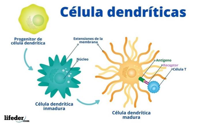
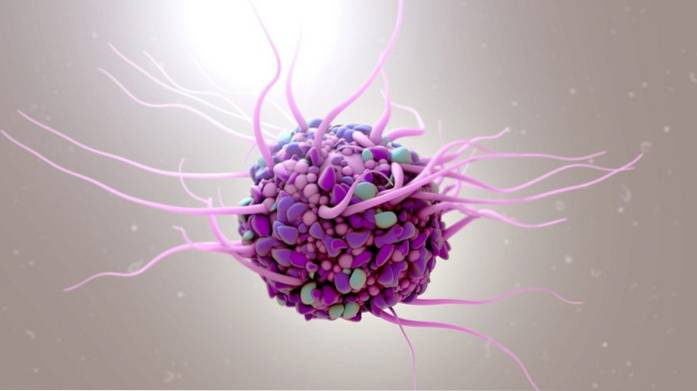
Dendritic cells are cells with an irregular appearance, which receive their name thanks to long extensions or projections of their plasma membrane that resemble the branches of a tree or the dendrites of nerve cells..
Both the shape and the ability to move dendritic cells depend on the stage of development and their function (antigen capture and presentation)..
They are found in the interstitium of many organs -except in the brain- and are particularly abundant in the regions of the body most exposed to the external environment, such as the skin, the mouth, the female genitalia, etc..
These cells differ from other cells by their high level of expression of a group of protein molecules known as the major histocompatibility complex class II or MHC II..
The proteins of this complex have much to do with the function of dendritic cells, since it is these that can bind to the antigens processed intracellularly to be presented to helper T lymphocytes in order to promote the activation of their immune functions..
Dendritic cells also present other surface markers, but these can vary depending on the type of cell, its stage of development and its location in the body..
Dendritic cells belong to the group of antigen-presenting cells of the immune system, where they are known as professional antigen-presenting cells..
These specialize in presenting antigens to other cells of the immune system known as helper T lymphocytes (helpers), which is why they function as messengers between the innate and adaptive immune systems.
Its main task is, then, to process the antigens derived from different classes of pathogens and expose them on their surface so that the cells of the immune system responsible for recognizing them and causing the immune response come into contact with said antigens..
To participate in the activation of the immune responses of the human body, dendritic cells must first come into contact with antigens derived from different sources, such as bacteria, fungi, viruses, parasites, etc., which they somehow achieve enter the body.
The contact and presentation of antigens are two events that are separated in time and space:
- Dendritic cells are found in many body tissues that are relatively exposed to the outside environment, especially in the dermis of the skin. It is in these tissues that they come into contact with the antigenic particles that are soluble or present on the surface of an invading pathogen, whom they must efficiently capture..
- When these antigens are internalized and internalized, signals are fired inside the dendritic cells that cause them to migrate to the nearest secondary lymphoid organ, where they differentiate at a stage of their development that allows the selection and presentation of antigens to the cells. helper T cells.
Dendritic cells not only activate helper T cells or lymphocytes that recognize the antigens that are presented to them, but also activate another group of effector cells known as cytotoxic T lymphocytes, capable of migrating to the site from which the dendritic cells migrated and eliminate those cells infected by the invading pathogen.
Dendritic cells form a relatively heterogeneous group of cells, both from the point of view of their origin as well as their functions and their surface markers. However, it is necessary to establish that these can be found in three different stages of development:
Now, a common classification of these cells assumes the existence of 4 groups, namely:
They are derived from precursors in the bone marrow and are part of the mononuclear phagocyte system. They have a restricted mitotic activity, so they are continuously replaced when they migrate towards the tissues where they lodge.
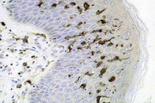
Langerhans cells are among the most studied; are very common in the dermis and in the epithelium of the oral cavity, esophagus and vagina.
They are cells with a dense nucleus, pale cytoplasm and membrane processes or extensions that radiate from the cell body to the intercellular spaces of the epidermal cells..
Like other cells in the human body, these cells have some mitochondria, a dispersed endoplasmic reticulum and some lysosomes, multivesicular bodies, and also many very small individual vesicles..
They are distinguished from the cells around them by the presence, in their membrane, of vermiform granules called Birbeck granules, that resemble miniature ping-pong rackets.
These granules contain a protein known as langerina, that participates in the internalization of antigens in the area, as well as in their degradation to the epitopes that later present, in the lymph nodes, to the T lymphocytes.
Langerhans cells are also characterized by the expression of a type of antigens known as CD1a, a group of surface proteins similar to those of the major histocompatibility complex that is responsible for presenting antigens such as peptides or other non-protein microbial antigens..
Found in most organs and body tissues, including the lungs, heart, kidneys, and dermis, they are important reservoirs for immature precursors of this cell type..
Unlike Langerhans cells, interstitial dendritic cells do not have Birbeck granules and do not always express CD1a antigens..
This group is also known as the "conventional dendritic cells". They are cells that when immature have a great phagocytic activity and when they mature they have a strong capacity to present antigens, as well as to secrete enormous amounts of cytokines.
Its surface markers are both the molecules of the major histocompatibility complex I and II (MHC I and III) and other molecules called CD11c, CD33 and CD13, which are markers of cells belonging to the myeloid lineage..
These cells are present in the circulatory system, but are also found in virtually any peripheral tissue and lymphoid organ in the body..
They are dendritic cells specifically associated with lymphoid tissues such as the tonsils, lymph nodes, and spleen. It is a set of migratory cells that belongs to the lymphoid lineage and not to the myeloid, like the previous group.
They present, on their surface, MHC type I and II markers, common leukocyte antigens and complement receptors. They are defined as accessory cells of the T-lymphocyte-dependent immune response.

Yet No Comments