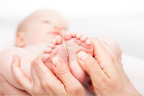
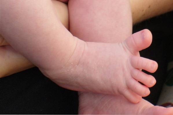
The reflection or Babinski's sign, Also known as the plantar reflex, it is used to know the degree of brain maturity or if there is any nervous pathology. It occurs when the sole of the foot is rubbed with a specific tool; the big toe moves up and the other toes fan out. Aims to protect the sole of the foot from possible damage.
This reflex is usually present in babies up to the age of two, approximately. In adults it is considered an abnormality, since it may indicate damage to the pyramidal pathway of the spinal cord, which is responsible for controlling voluntary movements.
If an older child or adult has this sign, it is possible that there is some neurological condition such as tumors in the spinal cord, strokes, multiple sclerosis, meningitis, etc..
Article index
The Babinski reflex was described by the French neurologist Joseph Françoise Félix Babinski in the late 19th century. This author was the first to report this phenomenon at a meeting of the Société de biologie in 1896.
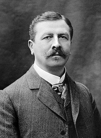
Babinski was looking for signs and reflexes that might distinguish organic from hysterical hemiparesis. During this period, several neurologists were trying to differentiate these two conditions. Thus, Babinski realized that this reflex could be related to some organic disturbances of the nervous system.
He also observed this reflex in patients with hemiplegia, a condition in which half of the body becomes paralyzed. In this way, he compared the response of the toes of the affected side with the response of the intact side, taking the healthy foot as control..
In another article on the subject published in 1898, Babinski highlighted the fact of the extension of the big toe during stimulation of the sole of the foot.
He analyzed the reflex in various clinical situations, without finding it in patients with hysterical weakness. In addition, he saw that it could be absent in people with hemiplegia or paraplegics with diminished, normal or absent myotatic reflexes (the one that occurs when a skeletal muscle is stretched).
In this way, he verified that the weakness of the reflex is not directly related to the intensity of the paralysis..
In 1903, Babinski published a last article. In it he described that this reflex was observed in patients who had alterations in the a pyramidal system or with congenital spastic paralysis. Also in newborns, in whom the nervous system has not fully developed.
The Babinski reflex in an adult, from the phylogenetic point of view, indicates a regression to a primary stage of development, where the locomotor system has not matured.
Doctors can elicit the Babinski reflex on a physical exam. To do this, the lateral part of the foot is rubbed with a flat instrument. This is specially designed not to cause pain, discomfort or injury to the skin.
Gentle pressure or caressing from any part of the leg could produce the reflex as well, but the most effective method is stimulation of the sole of the foot..
The instrument is passed from the heel forward, until it reaches the base of the toes. The Babinski reflex is clearly seen in newborns, as long as the surface is not very gently stimulated. Since, in this case, a grip reflex would occur.
Stimulation can elicit four different responses:
- Flexion: The toes are arranged downward and inward. The foot is placed in the eversion position (the bone that forms the heel moves away from the line that passes through the center of the body).
This is the response that occurs in healthy adults. It can be called "negative Babinski reflex".
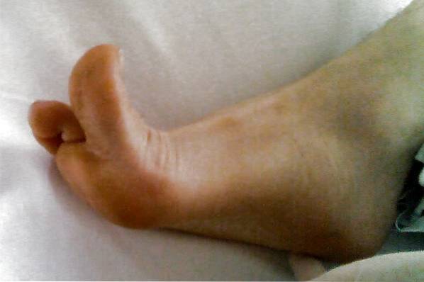
- Extension: there is a dorsiflexion of the big toe (approaching the shin) and the other toes are fanned out. This is the Babinski sign and is named as "positive Babinski reflex." It is observed in newborns, while in adults it implies some pathology.
- Indifferent: there's no answer.
- Ambiguous: there may be flexion of the toes before extension. Other times the flexor reflex can occur on one side, while the toe remains neutral on the other side..
In these cases, it is not clear whether there are lesions in the corticospinal tract. Therefore, other tests that are variants of the Babinski reflex should be performed..
The Babinski reflex can be tested in different ways. The usual way is the one explained in the previous point, since it seems to be the most reliable.
However, when ambiguous answers are given, the existence of the Babinski reflex can be corroborated using one of its variants.
- Schaefer's variant (1899): involves pinching the Achilles tendon enough to cause pain.
- Oppenheim's variant (1902): in this, strong pressure is applied with the thumb and index finger on the anterior part of the tibia up to the ankle.
- Gordon's variant (1904): it compresses the calf muscles by exerting deep pressure on them.
- The Chaddock variant (1911): It consists of stimulating the lateral malleolus (one of the bones that protrudes from the ankle) by hitting the skin around it, making circles. Can also be stimulated forward, from heel to little toe.
- Variant of Bing (1915): the back of the big toe is pricked with a pin. A pathological reaction would be for the finger to extend upward toward the pin. While a normal reaction would be to flex the finger downward, fleeing from the puncture.
This last sign, together with Chaddock's, are the most reliable after Babinski's sign..
The plantar reflex has been understood to involve more movements than just the toes. In most mammals, the limbs automatically retract on a painful stimulus. This defensive reflex is controlled by polysynaptic pathways in the spinal cord..
The reaction is more pronounced in the hindlimbs, since the forelimbs are under more direct control of the brain. Not only the skin, but deeper structures have receptors that can generate this movement.
The reflex effects on the human leg when stimulating the sole of the foot are comparable to those of animals.
Most newborns and young children are not neurologically mature, thus showing the Babinski reflex. Unlike the older ones, in babies the flexion is much faster. The toes rise at the same time as the ankle, knee and hip flexion..
As the pyramidal system matures and there is more control of the spinal motor neurons, there are changes in the flexion reflex. The most important change occurs after one or two years, and is that the fingers are no longer part of the flexion synergy.
While another observed change is that the flexion reflex becomes less pronounced.
However, the neurophysiology of the Babinski reflex is not yet fully understood. From electromyographic studies, it is known that each area of skin appears to have a specific reflex response to noxious stimuli. The purpose of the reflex is to provoke the withdrawal of the skin of such stimulus.
The area of the skin from which the reflex can be obtained is called the "reflex receptive field." Specifically, when there is a noxious stimulus on the sole of the foot (which would be a receptive field) the body reacts.
There is an immediate flexion of the toes, ankle, knee and hip joints, away from the stimulus. This is what happens when we step on a sharp object with bare feet. There is an involuntary flexion of all the joints and the withdrawal of the foot.
Another normal individual reflex is the big toe reflex. Stimulation of the receptive field of the ball of the foot causes extension of the toe, in addition to flexion of the ankle, knee and hip joints.
The difference between these two types of reflections is in the receptive fields. It is the reason why in one the big toe is flexed and in another it is extended.
What happens in the Babinski reflex is that an extension of the big toe occurs when the wrong receptive field is stimulated. Therefore, in the face of a noxious stimulus to the sole of the foot, the toe is extended instead of the normal flexion response..
In newborns and infants up to two years of age, the central nervous system is not fully developed. In this way, there are parts of the corticospinal tract still without myelin (layers that cover neurons and that facilitate the transmission of information).
The corticospinal tract or pyramidal tract are very long nerve axons. They originate in the cerebral cortex, and go from the brainstem to the spinal cord. The neurons of the corticospinal tract are known as "upper motor neurons".
The cortiospinal tract influences the spinal cord reflex. When this tract is not working properly, the receptive field of the reflex increases to encompass a different receptive field..
It seems that the adequate conservation of receptive fields depends on an intact cerebral cortex..
An abnormal Babinski reflex may be the first indication of serious disease, so more detailed tests such as a CT scan, MRI, or lumbar puncture should be performed to study the cerebrospinal fluid..
Under normal conditions, the Babinski reflex would be present in children less than two or three years of age. And from this age on, it would disappear and be replaced by the flexor reflex.
If this reflex does not appear in the first 6 months of age, this is known by some authors as a negative Babinski reflex. This could mean that there are neurological abnormalities such as cerebral palsy, mental retardation; or less frequent, motor lag. (Futagi, Suzuki & Goto, 1999).
The Babinski reflex in adults or older children reliably indicates that there is a metabolic or structural abnormality in the corticospinal system.
This can be manifested by symptoms such as lack of coordination, weakness, and difficulty controlling muscle movements..
It is also pathological to have the Babinski reflex on one side of the body, but not on the other. This could suggest which side of the brain is affected.
On the other hand, an abnormal Babinski's sign can be temporary or permanent, depending on the condition causing it..
Some of the conditions associated with this reflex are:
- Spinal cord injury or tumors.
- Syringomyelia or spinal cysts.
- Meningitis: is a disease in which there is severe inflammation of the membranes that cover the brain and spinal cord.
- Stroke or stroke.
- Amyotrophic lateral sclerosis (ALS): consists of a degenerative neurological disease that affects the motor neurons of the brain or spinal cord.
- Friedreich's ataxia: is a neurodegenerative condition that causes deterioration in the cerebellum and dorsal spinal ganglia.
- Poliomyelitis: consists of an infection that attacks the spinal cord, causing muscle atrophy and paralysis.
- Brain tumor or damage involving the corticospinal tract.
- Abnormal metabolic states such as hypoglycemia (low blood glucose), hypoxia (lack of oxygen), and anesthesia.
- Multiple sclerosis: it is a degenerative condition of the central nervous system. Progressive brain and spinal cord injuries occur. It is possible that an abnormal Babinski reflex may indicate multiple sclerosis, although not all people with multiple sclerosis have this reflex..
- Pernicious anemia: infection characterized by insufficient red blood cells, which are responsible for providing oxygen to the body's tissues.
- After experiencing generalized tonic-clonic seizures.

Yet No Comments